

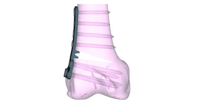
If you’ve been told you have valgus alignment, or “knock-knee” alignment, you may be experiencing pain on the outside of your knee that worsens with activity. In this condition, your leg alignment places extra stress on the outer part of the knee. Over time, this increased pressure can damage the cartilage and meniscus, leading to pain, swelling, and early arthritis. For many younger, active patients, a distal femoral osteotomy (DFO) can be a life changing operation.
Valgus malalignment is usually diagnosed with full-length alignment films. The doctor will draw mechanical axis (a line from the middle of the femoral head to the middle of the ankle). This line should generally fall near the middle of the knee. However, in those with valgus or knock knee alignment, the line falls along the lateral our outside portion of the knee (Figure 1).
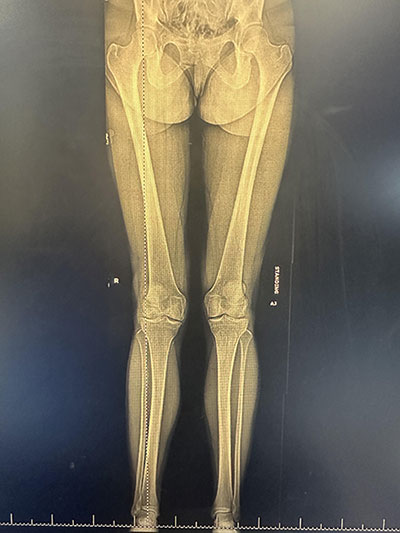
Figure 1
A distal femoral osteotomy is a procedure that corrects the alignment of the leg by making a carefully planned cut in the lower part of the thigh bone (femur). The distal femur is cut with a saw blade and the cut in the bone is opened up with gentle manual pressure. Once the gap is appropriate based on mathematical measurements, a plate with screws is applied to hold the osteotomy in place. The bone is repositioned to shift your body weight away from the damaged area of the knee and more so through the middle of the knee (Figure 2). By restoring a more normal alignment, the procedure can reduce pain, improve function, and help protect the knee from further damage.
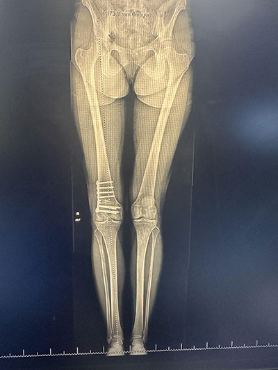
Figure 2
Not everyone with knee pain is a candidate for a DFO. This procedure is best suited for patients who are generally younger and active, have pain primarily on the outside of the knee, and have damage that is limited to one part of the joint. Many patients have also experienced a meniscus injury or cartilage damage that contributes to their symptoms.
A DFO is often performed along with other procedures to improve the overall health of the knee. These may include cartilage restoration surgery, meniscus transplantation, or ligament reconstruction when needed. Whenever one performs significant cartilage work, such as an osteochondral allograft, or meniscal work (significant meniscal repair or meniscal transplant), one should always check alignment in the clinic prior to undergoing such an operation. Performing cartilage or meniscal preservation without fixing alignment can be a recipe for poor outcomes and the need for revision surgery.
Our clinic routinely obtains a CT scan of the lower extremity to order patient specific instrumentation guides to help with cutting and custom plates when performing osteotomy fixation. Studies have shown that custom guides and plates can help with the accuracy of planned corrections while reducing operative time and radiation exposure.
Recovery takes time and commitment. Most patients use crutches for several weeks while the bone heals and participate in a structured physical therapy program to restore motion, strength, and balance. Although recovery is gradual, many patients are able to return to the activities they enjoy once healing is complete.
One of the biggest advantages of a distal femoral osteotomy is that it preserves your own knee. Rather than replacing the joint, the surgery corrects the underlying problem that caused the abnormal wear in the first place. For many patients, this can delay the need for a knee replacement for many years. Studies have shown good to excellent clinical outcomes.
Every patient is different, which is why careful evaluation is so important. Standing alignment X-rays, advanced imaging, and a thorough physical examination help determine whether a DFO is the right treatment option.
If you’re experiencing persistent pain on the outside of your knee and have been told you have knock-knee alignment or early arthritis, a distal femoral osteotomy may be worth discussing with your orthopedic surgeon. When performed in the right patient, this procedure can relieve pain, improve function, and help you stay active while preserving your natural knee.
Amit Momaya, MD is a board certified orthopedic surgeon who specializes in sports medicine. He serves as the Chief of Sports Medicine at UAB, resides on the editorial board of research journals, and takes care of several collegiate and professional teams in Alabama. This blog post is for educational purposes only. Please consult with your physician for a discussion on knee preservation.


